I thought about how many urethral complications there are with phallo and sought out some imaging studies. a urethrography involves putting a contrast into the urethra to get better images of the shape of it. in patients without suprapubic (above the pubic bone) catheters, they squirt constrast in from the outside. When they DO have a suprapubic catheter it usually makes more sense to do a voiding study, so they put contrast in through the catheter and take images while the patient pees. there are a mix of the voiding imaging studies and the normal kind.
anyway, I got a pic of a normal man having the same imaging study for reference;
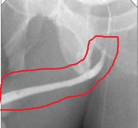
I circled the urethra in red. its just a smooth tube, exactly how it is supposed to be. the fainter structures behind that are bone and you can see the outline of thighs. the guys weiner is pointing to the left side of the picture, which is the case in all of these pics I included in the thread (one or two at the link are front facing and say so)
archived 6 May 2022 15:02:39 UTC

archive.is
here are some of the things I found from studies on phallo patients:
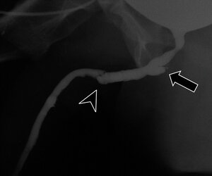
the surgery has deformed the natal urethral anatomy. maybe its like if you narrow a garden hose it may swell and burst further up the line? doesn't look good, in any case. the description notes that the "horns" (the structure on the right hand side w/an arrow) may persist indefinitely. if this patient ever gets a UTI this structure could act a reservoir for bacteria. what I am saying is true of every single pocket type structure you're going to see in the thread.
this next picture says "contained leak", which seems to mean that the pee leaks into pockets in subcutaneous tissue (so not into another organ). the arrows are gesturing towards the leaking contrast/urine mixture detected by a voiding urethrography (so they put contrast in through a catheter above the pubic bone, and then have the patient pee during imaging to see how the contrast moves)
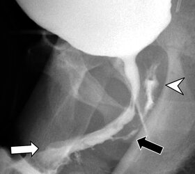
another contained leak :
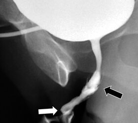
^^^this one references "horns", it is the shape of the leak. I don't know enough about urologic pathology to know if that is a normal shape for a urethral leak or if this is a problem that is common enough to phalloplasty to name it this way, but a lot of "horns" references throughout the paper.
a worse contained leak with many chambers filling with pee :
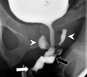
another contained leak that looks like a potato:
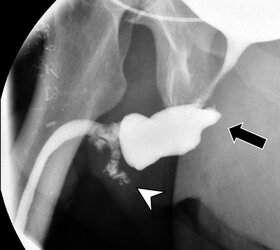
and now an anastomatic leak. anastomatic means the leak is between two organ structures. In this case the leak goes into a remnant of the vagina that the patient had after vaginectomy:
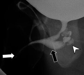
another contained leak, looks like some small ones along the neophallus itself:
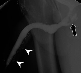
and now onto occlusions (blocking off of a structure) and stenosis (narrowing of a tube)
this is a complete occlusion, meaning no pee comes out. I suppose they must have done this via a suprapubic catheter, though I wonder how the fuck they get the contrast out after that.
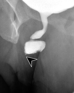
stenosis is pretty severe in this patient, the arrow is pointed at the narrowing structure. if the narrowing is a result of scarring then you can generally expect it to get worse over time. the treatment of stenosis is mechanical dilation.
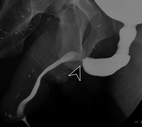
this is a case with contained leaks and stenosis:
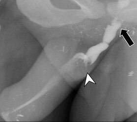
^^this one almost looks like the bulges in the urethra are creating a telescoping structure. In anatomy this is called intussusception but that condition is mostly associated with the intestines, and it is a pretty serious problem. I hadn't heard of this happening to urinary structures but it has apparently happened in the past once or twice with natal urethras and many more times with the ureters (the tubes that go from the kidneys to the bladder). Intussusception is a problem because it can cut off blood flow to the areas squeezed by the telescoping action.
here are two images that have fistulas and stenosis:
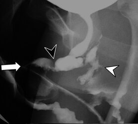
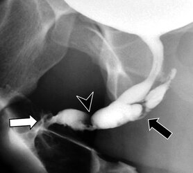
these are presumably the same patient before and after surgical treatment of the fistulas. the stenosis wasn't treated because it apparently wasn't causing symptoms, though to me it seems like the stenosis likely caused the pressure that formed the fistula. I'm not a urologist though so idk, not that any of this is established well enough through science for any urologist to know much about what the fuck they are doing, either.
delayed fistula:
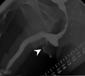
looks like the fistula goes out of the body under the neophallus, presumably dribbles pee everywhere.
I left out a couple of the pictures in the article but feel free to look them over.
it really is kind of psychopathic to assume that you can use skin or buccal (inside of the cheek) tissue to replace urethra. I know that buccal tissue can be used as a patch on many mucus membrane repairs, but this is like if you tried to make a garden hose by sewing together hose patches and then affixed them to the end of a normal hose. it isn't likely to work the same way as a discrete structure. many of these pics have coke can dick, too.
you remember those laws that made women hear the fetal heartbeat before they got abortions? If I were to write a law, it would be like that except SRS patients would have to see this type of shit and understand the pictures before agreeing to surgery. Then we wouldn't have to feel bad for any of them anymore and presumably those with any sense would escape this fate.
![lightbox_d44a9280f77611eab7c6377b387e4fef-Figure-1-resized[1].png lightbox_d44a9280f77611eab7c6377b387e4fef-Figure-1-resized[1].png](http://uploads.kiwifarms.st/data/attachments/3250/3250332-e310b4b522f7610eec2c1b4b3f429177.jpg?hash=4xC0tSL3YQ)


